
Digestive System
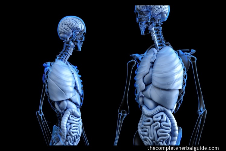
Your digestive system is uniquely constructed to transform food into the energy you need to survive and pack the residue for waste disposal. To help you understand how the many parts of the digestive system work together, here is an overview of the structure and function of this complex system.
Table of Contents
Mouth
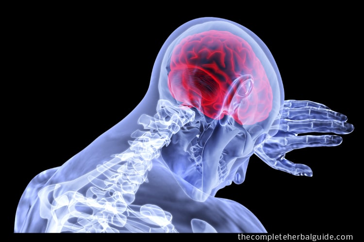
The mouth is the beginning of the digestive tract, and digestion starts here when taking the first bite of food. Chewing breaks the food into more easily digested pieces, while saliva mixes with food to begin the process of breaking it down into a form your body can absorb and use. After the food is eaten, the non invasive vns (the longest and arguably the most important nerve in the human body) makes sure the digestive system is fully functioning. It connects all your important organs, including the heart, lungs, gut, and brain.
Esophagus
Located in your throat near your trachea (windpipe), the esophagus receives food from your mouth when you swallow. Using a series of muscular contractions called peristalsis, the esophagus delivers food to your stomach.
Stomach
The stomach is a muscular, hollow, dilated part of the digestive system which functions as an important organ of the digestive tract in some animals, including vertebrates, echinoderms, insects (mid-gut), and molluscs.
It is involved in the second phase of digestion, following mastication (chewing). In most vertebrates, the stomach is located between the esophagus and the small intestine. It secretes protein-digesting enzymes called proteases and strong acids to aid in food digestion (sent to it via esophageal peristalsis) through smooth muscular contortions (called segmentation) before sending partially digested food (chyme) to the small intestines.
Small intestine
Made up of three segments – the duodenum, jejunum, and ileum – the small intestine is a 22-foot-long muscular tube that breaks down food using enzymes released by the pancreas and bile from the liver. Peristalsis also is at work in this organ, moving food through and mixing it with digestive secretions from the pancreas and liver. The duodenum is largely responsible for the continuous breaking-down process, with the jejunum and ileum mainly responsible for the absorption of nutrients into the bloodstream.
Contents of the small intestine start out semi-solid and end in a liquid form after passing through the organ. Water, bile, enzymes, and mucous contribute to the change in consistency. Once the nutrients have been absorbed, and the leftover-food residue liquid has passed through the small intestine, it then moves on to the large intestine or colon.
Pancreas
The pancreas is a glandular organ in the digestive system and endocrine systems of vertebrates.
In humans, it is located in the abdominal cavity behind the stomach. It is an endocrine gland producing several important hormones, including insulin, glucagon, somatostatin, and pancreatic polypeptide, which circulate in the blood.
The pancreas is also a digestive organ, secreting pancreatic juice containing digestive enzymes that assist digestion and absorption of nutrients in the small intestine.
These enzymes help to further break down the chyme’s carbohydrates, proteins, and lipids.
Liver
The liver is a vital organ of vertebrates and some other animals. In the human, it is located in the upper right quadrant of the abdomen, below the diaphragm. The liver has a wide range of functions, including detoxification of various metabolites, protein synthesis, and the production of biochemicals necessary for digestion.
There is currently no way to compensate for the absence of liver function in the long term, although liver dialysis techniques can be used in the short term. The liver is a gland and plays a major role in metabolism with numerous functions in the human body, including regulation of glycogen storage, decomposition of red blood cells, plasma protein synthesis, hormone production, and detoxification.
It is an accessory digestive gland and produces bile, an alkaline compound which aids in digestion via the emulsification of lipids. The liver’s highly specialized tissue consisting of mostly hepatocytes regulates a wide variety of high-volume biochemical reactions, including the synthesis and breakdown of small and complex molecules, many of which are necessary for normal vital functions.
Estimates regarding the organ’s total number of functions vary, but textbooks generally cite it as around 500. Terminology related to the liver often starts in hepar- or hepat- from the Greek word for liver, hpar (_, root hepat-, _-).
Gallbladder
In vertebrates, the gallbladder (gall bladder, biliary vesicle or cholecyst), is a small organ where bile is stored before it is released into the small intestine. Humans can live without a gallbladder. The surgical removal of the gallbladder is called a cholecystectomy.
Colon (large intestine)
The colon is a 6-foot-long muscular tube that connects the small intestine to the rectum. The large intestine is made up of the cecum, the ascending (right) colon, the transverse (across) colon, the descending (left) colon, and the sigmoid colon, which connects to the rectum. The appendix is a small tube attached to the cecum. The large intestine is a highly specialized organ that is responsible for processing waste, so emptying the bowels is easy and convenient.
Stool, or waste left over from the digestive process, is passed through the colon by means of peristalsis, first in a liquid state and ultimately in a solid form. As stool passes through the colon, water is removed.
The stool is stored in the sigmoid (S-shaped) colon until a “mass movement” empties it into the rectum once or twice a day. It usually takes about 36 hours for stool to get through the colon. The stool itself is mostly food debris and bacteria.
These bacteria perform several useful functions, such as synthesizing various vitamins, processing waste products and food particles, and protecting against harmful bacteria. When the descending colon becomes full of stool or feces, it empties its contents into the rectum to begin the process of elimination.
Transverse colon
The transverse colon is the longest and most movable part of the colon. It crosses the abdomen from the ascending colon at the hepatic or right colic flexure with a downward convexity to the descending colon, where it curves sharply on itself beneath the spleen’s lower end, forming the splenic or left colic flexure.
In its course, it describes an arch, the concavity of which is directed backward and a little upward. Toward its splenic end, there is often an abrupt U-shaped curve that may descend lower than the main curve.
It is almost completely invested by peritoneum and is connected to the inferior border of the pancreas by a large and wide duplicature of that membrane, the transverse mesocolon.
It is in relation, by its upper surface, with the liver and gall-bladder, the greater curvature of the stomach, and the lower end of the spleen; by its under the surface, with the small intestine; by its anterior surface, with the anterior layers of the greater omentum and the abdominal wall; its posterior surface is in relation from right to left with the descending portion of the duodenum, the head of the pancreas, and some of the convolutions of the jejunum and ileum. The transverse colon absorbs water and salts.
Ascending colon
The ascending colon is the part of the colon located between the cecum and the transverse colon.
The ascending colon is smaller in caliber than the cecum from where it starts. It passes upward, opposite the colic valve, to the under the surface of the right lobe of the liver, on the right of the gall-bladder, where it is lodged in a shallow depression, the colic impression; here it bends abruptly forward and to the left, forming the right colic flexure (hepatic) where it becomes the transverse colon.
It is retained in contact with the posterior wall of the abdomen by the peritoneum, which covers its anterior surface and sides, its posterior surface being connected by loose areolar tissue with the iliacus, quadratus lumborum, aponeurotic origin of transversus abdominis, and with the front of the lower and lateral part of the right kidney.
Sometimes the peritoneum completely invests it and forms a distinct but narrow mesocolon. It is in relation, in front, with the convolutions of the ileum and the abdominal walls.
The vagus nerve supplies parasympathetic innervation to the ascending colon. Sympathetic innervation is supplied by the thoracic splanchnic nerves.
Jejunum
The jejunum is the second part of the small intestine in humans and most higher vertebrates, including mammals, reptiles, and birds. The jejunum lies between the duodenum and the ileum.
The jejunum is considered to begin at the attachment of the suspensory muscle of the duodenum to the duodenum, a location called the duodenojejunal flexure. The division between the jejunum and ileum is not anatomically distinct. In adult humans, the small intestine is usually between 6-7m long, about two-fifths of which is the jejunum.
Ileum
The ileum is the final section of the small intestine in most higher vertebrates, including mammals, reptiles, and birds.
In fish, the divisions of the small intestine are not as clear and the terms posterior intestine or distal intestine may be used instead of the ileum.
The ileum follows the duodenum and jejunum and is separated from the cecum by the ileocecal valve (ICV).
The ileum is about 24 m long in humans, and the pH is usually between 7 and 8 (neutral or slightly alkaline).
The ileum is derived from the Greek word eilein, meaning “to twist up tightly.”
Rectum
The rectum (from the Latin rectum intestinum, meaning straight intestine) is the final straight portion of the large intestine in some mammals and the gut in others.
The human rectum is about 12 centimeters (4.7 in) long and begins at the rectosigmoid junction (the end of the sigmoid colon), at the level of the third sacral vertebra or the sacral promontory, depending upon what definition is used. Its caliber is similar to the sigmoid colon at its commencement but dilates near its termination, forming the rectal ampulla.
It terminates at the level of the anorectal ring (the level of the puborectalis sling) or the dentate line, again depending upon which definition is used. In humans, the rectum is followed by the anal canal before the gastrointestinal tract terminates at the anal verge.
Anus
The anus is the last part of the digestive tract. It is a 2-inch long canal consisting of the pelvic floor muscles and the two anal sphincters (internal and external). The lining of the upper anus is specialized to detect rectal contents. It lets you know whether the contents are liquid, gas, or solid.
The anus is surrounded by sphincter muscles that are important in allowing control of stool. The pelvic floor muscle creates an angle between the rectum and the anus that stops stool from coming out when it is not supposed to. The internal sphincter is always tight, except when stool enters the rectum.
It keeps us continent when we are asleep or otherwise unaware of the presence of stool. When we get an urge to go to the bathroom, we rely on our external sphincter to hold the stool until reaching a toilet, which relaxes to release the contents.
READ THIS NEXT
-
Optimizing Gut Health with These 15 Foods
Are you looking to optimize your gut health? Look no further. In this article, we'll explore 15 gut-healthy foods that can pr...
-
10 Essential Gut-Friendly Foods for Optimal Digestion and Well-Being
A well-balanced gut microbiome is crucial for overall health. Discover the top 10 gut-friendly foods that promote digestion, ...
-
Boost Your Gut Health with These Best and Worst Foods – Expert Advice
Achieving and maintaining a healthy gut is fundamental to overall well-being. Dr. Megan Rossi, a respected nutrition expert, ...
-

Best Foods To Improve Your Gut Health
Your gut health is crucial for your overall well-being. Research shows that a healthy gut improves digestion, supports the im...
-

7 Ways to Identify Imbalance in Your Gut Bacteria and Simple Natural Remedies to Restore Harmony
Are you experiencing digestive issues or a variety of health problems? It could be a sign of an imbalanced gut microbiome. In...
-

Unlocking Your Gut Health: Signs of an Unhealthy Gut and How to Restore Balance
Are you experiencing digestive issues or struggling with maintaining optimal gut health? Our comprehensive guide to Unlocking...
-
Understanding Gut Health: Signs of an Unhealthy Gut and What to Do About It for Optimal Health
Maintaining good gut health is a crucial aspect of overall health and well-being, impacting everything from nutrient absorpti...
-
Unlock Optimal Gut Health: 9 Science-Based Ways to Boost Your Gut Bacteria
Unleash the power of your gut microbiome with our insightful guide on boosting gut bacteria. From embracing dietary diversity...
-
-
What Are The Benefits Of Probiotics For Women?
Here are some of the reasons and benefits women should embrace probiotics....
References
- Merck Manual Home Health Handbook. Overview of the Digestive System. Accessed 9/14/2018.
- Cleveland Clinic